
No matter the amount of rigorous training and conditioning that goes into an athlete’s regimen, sports injuries are often an unfortunate and unavoidable part of the deal. Fortunately, radiological imaging has changed the way sports injuries have been diagnosed and treated over the years, allowing athletes to find a safer road to recovery, and a sure path back to the field in most cases.
The responsibility of accurate diagnosis from the start is vital. In sport, an athlete’s entire career is often dependant on the decisions made immediately following the injury. Radiologists are thus acutely aware of the responsibility that lies with them when included in the diagnosis.
With the various forms of radiology available, it may be necessary to look at the different imaging modalities and their specific benefits and uses when it comes to sports injuries.
X-rays
In the occurrence of an on-field injury where the possibility of a fracture or dislocation exists, X-rays offer the best imaging solution due to its common availability and quick turnaround time. X-rays are often relied on even if only to rule out the possibility of bone fracture or displacement.
Ultrasonography
Advancements in ultrasonographic technology have allowed imaging to display muscular architecture at less than 0.1mm in-pane resolution. By revealing cortical discontinuity, ultrasound is also able to identify subtle fractures not picked up by X-rays. This imaging modality is also able to indicate fibre disruption in muscles, tendons, and nerves before and during treatment, helping to ensure a holistic treatment approach throughout the process.
Computed tomography scanning (CT Scans)
While CT scans may offer slightly inferior soft tissue imaging compared to MRI scans, they are often more accessible and may be preferable in sporting injuries that involve possible bone damage since it has a quicker examination time. CT scans are often also the chosen modality for post-operative imaging procedures.
Image-guided injections
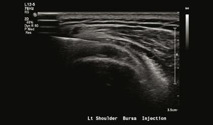
A major benefit of image modalities lies in the guidance of musculoskeletal injections that were previously delivered “blindly” through the guidance of clinical skills and anatomical landmarks alone. Thanks to advancements in radiology, ultrasound and CT-guided injections have become the norm in many injection procedures. Image guidance has also been of great benefit in the removal of collections, haematoma, and joint fluid that are often caused by sports injuries.
Magnetic resonance imaging (MRI)
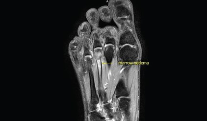
MRIs are a staple in the diagnosis and treatment of musculoskeletal injuries due to its ability to show soft tissue contrast and multiplanar visibility. This is of primary benefit to athletes in MRI’s ability to indicate bone marrow and soft tissue oedema, as well as to create imaging of the muscles, tendons, and ligaments. MRIs allow radiologists and medical practitioners to identify the active cause of pain.
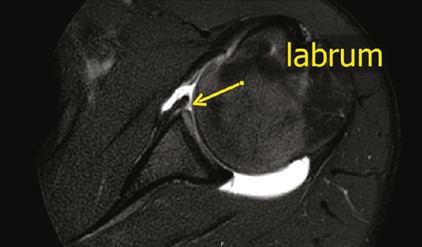
MRIs, are primarily relied upon following X-ray and ultrasonography findings, though, and especially valuable in determining a return to the sports arena post-injury. As such, they play a vital part in many athletes’ long-term health assurance.
Cartilage imaging
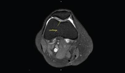
In the occurrence of blunt trauma to joints, especially the knees, MRIs have become highly valuable thanks to their effective evaluation of knee pain through the creation of accurate measurements of the cartilage ultrastructure.
Physeal fractures
Especially in adolescent athletes, physeal injuries, where the growth plate of the patient is fractured, a T1-weighted sequence MRI will help to evaluate even the slightest damage to the bone structure.
Peripheral nerve damage
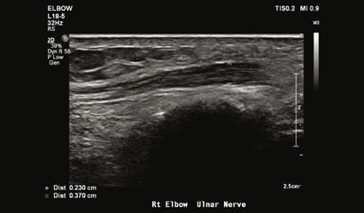
Sports injuries are most often related to muscle, tendon, and ligament injuries, and so, imaging is focused on soft tissue. However, peripheral nerve damage may also be a concern. Both MRIs and high-frequency sonography are able to depict these peripheral nerves and can help lead more effective treatment.
Acute versus chronic injury
Due to MRI’s sensitivity to the presence of oedema, it is able to help medical practitioners differentiate between chronic and astute pain by assessing scarring and thickening that are detectible in ligaments where repetitive stress is present.
Fortunately, just as with many of the sports players they treat, radiologists are also part of a professional team that consists of sports physicians, general practitioners, physiotherapists and Orthopedic surgeons, who all work together to get an athlete back on the field again, fully recovered. Being part of both the identification of the injury as well as the treatment process, they are key players present throughout gameplay, from the first whistle to the last goal.
Radiology forms a constant part of any athlete’s recovery, much like the playbook will form a part of a team’s next gameplay. Initial diagnoses and treatments are often adjusted in the course of recovery thanks to the active participation of the radiological department that identifies the effect and progress of treatment quicker and more accurately than external examination through internal imaging.
So, when the time comes to choose your team for a sport’s related injury, make sure to make an experienced radiologist one of your first picks.
This article is a general information sheet and should not be used or relied on as professional advice. No liability can be accepted for any errors or omissions nor for any loss or damage arising from reliance upon any information herein. Errors and omissions excepted (E&OE).