
We have your breast interests at heart
What you need to know about Mammography.

Mammography is a term for the X-Ray imaging of breasts. A mammogram is an X-ray image of your breasts used to detect tumours and other abnormalities and screen for breast cancer. Mammograms play a key role in early breast cancer detection and help decrease breast cancer deaths.
During a mammogram, your breasts are compressed between two firm surfaces to spread out the breast tissue. Then an X-ray captures black-and-white images of your breasts that are displayed on a computer screen and examined by a doctor who looks for signs of cancer.
A 3D mammogram is used as a breast cancer screening test to look for breast cancer in people with no signs or symptoms of the disease. It can also be used to investigate breast problems, such as a suspicious lump or thickening. While 3D mammography has a wide array of benefits, it is not necessarily recommended above standard 2D mammography (apart from a few specific instances).
Mammography is a term for the X-Ray imaging of breasts. A mammogram is an X-ray image of your breasts used to detect tumours and other abnormalities and screen for breast cancer. Mammograms play a key role in early breast cancer detection and help decrease breast cancer deaths.
During a mammogram, your breasts are compressed between two firm surfaces to spread out the breast tissue. Then an X-ray captures black-and-white images of your breasts that are displayed on a computer screen and examined by a doctor who looks for signs of cancer.
A 3D mammogram is used as a breast cancer screening test to look for breast cancer in people with no signs or symptoms of the disease. It can also be used to investigate breast problems, such as a suspicious lump or thickening. While 3D mammography has a wide array of benefits, it is not necessarily recommended above standard 2D mammography (apart from a few specific instances).
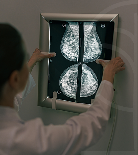
A mammogram can be used either for screening or for diagnostic purposes. At a certain age, women will need to start having regular mammography tests to help detect early signs of breast cancer and to ensure their own safety. How often you should have a mammogram depends on your age and your risk of breast cancer.
Is used to detect breast changes in women who have no signs or symptoms or new breast abnormalities. The goal is to detect cancer before clinical signs are noticeable.
Is used to investigate suspicious breast changes, such as a new breast lump, breast pain, an unusual skin appearance, nipple thickening or nipple discharge. It is also used to evaluate abnormal findings on a screening mammogram. A diagnostic mammogram includes additional mammogram images.
There is no ideal age to start screening for breast cancer. Experts and medical organisations are divided on when women should begin regular mammograms and how often these tests should be performed. Mammograms before the age of 40 are usually uncommon unless you have a high risk of developing breast cancer. Mammogram tests can often be recommended on an annual or biennial basis.
The best course of action would be to talk with your doctor about your risk factors, your preferences, and the benefits and risks of screening. Together, you can decide what screening mammography schedule is best for you.
Our team of health professionals have been chosen for their ability and expertise.
We are committed to developing each of our team members beyond their current level of expertise. In this way we provide a consistent, quality service that patients can rely on.
In all our practises, we make our patients’ safety our top priority. This is why we will prepare you for every diagnostic test before we conduct it.
As we work with complex machines to carry out our imaging procedures, there are some inherent risks that each procedure presents, these are risks we want to minimise by putting your safety first.
Knowledge is the foundation of the work we do in the practise. We believe that, in order to provide you with the quality of service you deserve as patient, developing our expertise and proficiency in a variety of fields is a necessity.
We are committed to continue learning and broadening the scope of what we understand about radiology and its practise so that we can create a safer, positive experience for all our patients.
Few. Mammograms will expose you to low-dose radiation, but the dose is very low, and for most women the benefits of regular mammograms outweigh the risks posed by this amount of radiation.

It is best to schedule the test for a time when your breasts are least likely to be tender. This is often the week before or after your menstrual period.
Prepare and bring along your prior mammogram images so that the radiologist can compare them to your new images.
Do not use deodorant, roll-on, or powder before your mammogram. Metallic particles in various aerosols, powders and creams can interfere with the imaging.
It is preferable that you wear a two-piece outfit on the day of the test.
Consider over-the-counter pain medication if you find that having a mammogram is uncomfortable. Taking an over-the-counter pain medication, such as aspirin, acetaminophen (Tylenol, others) or ibuprofen (Advil, Motrin IB, others), about an hour before your mammogram might ease the discomfort of the test.
You will be given a gown and asked to remove neck jewellery and clothing from the waist up.
For the procedure itself, you will stand in front of an X-ray machine specially designed for mammography. The technician will place one of your breasts on a platform and raise or lower the platform to match your height. The mammographer will help you position your head, arms and torso to allow an unobstructed view of your breast.
Your breast is then gradually pressed against the platform by a clear plastic plate. Pressure is applied for a few seconds to spread out the breast tissue. The pressure isn’t harmful, but you may find it uncomfortable or even painful. We do, for this reason, offer the option of self-compression.
If you have too much discomfort, tell the technician. Your breast must be compressed to even out its thickness and permit the X-rays to penetrate the breast tissue. The pressure also holds your breast still to decrease blurring from movement and minimises the dose of radiation needed. During the brief X-ray exposure, you will be asked to stand still and hold your breath.
After images are made of both your breasts, you may be asked to wait while the technician checks the quality of the images. If the views are inadequate for technical reasons, you may have to repeat part of the test. The entire procedure usually takes less than 30 minutes. Afterward, you may dress and resume normal activity.
Your mammograms will appear on a computer screen. A radiologist interprets the images and sends a written report of the findings to your doctor. The radiologist looks for evidence of cancer or non-cancerous (benign) conditions that may require further testing, follow-up or treatment. If your breast tissue proves to be dense further testing may also be required.
If the radiologist notes areas of concern on your mammogram, further testing may include additional mammograms, known as compression or magnification views, as well as ultrasound imaging or a biopsy to remove a sample of breast tissue for laboratory testing.
Some situations require the use of diagnostic magnetic resonance imaging (MRI) in areas where the current imaging with mammography or ultrasound is negative and it is not clear what is causing a breast change or abnormality.
Shelly Beach Details
Monday – Sunday
Open 24/7
Amanzimtoti Details
Monday – Friday
8:00AM – 5:00PM
Shelly Beach Private Hospital
Umbogintwini, Amanzimtoti, Durban